What is Diagnostic Radiology (or Diagnostic Imaging)?
- Francesco Bruno Tagliaferro
- Jul 14, 2023
- 4 min read
Updated: Jul 25, 2023
Diagnostic radiology, or diagnostic imaging, is a branch of medicine that deals with the diagnosis and evaluation of disease through the creation, reading and interpretation of radiological images.
Radiodiagnostics includes several imaging techniques, including:

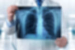
Radiography: uses X-rays to produce two-dimensional images of the body, allowing for visualization of bones, organs, and soft tissue.

Computed tomography (CT): combines X-rays and computer processing to create cross-sectional images of the body. CT provides detailed images of organs, blood vessels, soft tissue, and anatomical structures in several planes.

Magnetic resonance imaging (MRI): uses magnetic fields and radio waves to generate detailed images of organs and soft tissue. MRI offers great insight into the structure and function of the body, and is especially useful for visualizing the brain, spinal cord, joints, and soft tissue.

Ultrasound: uses ultrasound to generate real-time images of internal organs, blood vessels, and soft tissue. Ultrasound is widely used to examine the abdomen, heart, thyroid gland and other areas of the body.
Diagnostic imaging plays a vital role in the diagnosis and monitoring of numerous medical conditions, including traumatic injuries, cardiovascular disease, cancer, nervous system diseases, musculoskeletal disorders, and many others. Radiological images, together with the expertise of radiologists in interpreting these images, provide essential information for accurate diagnosis and appropriate treatment planning.
Radiology has had a huge impact on medicine and has turned into an indispensable specialty in modern medical practice for several reasons:
Early and accurate diagnosis. Radiology offers advanced imaging tools that allow for the early identification and diagnosis of a wide range of medical conditions. X-ray images, computed tomography (CT), magnetic resonance (MRI) and other techniques allow doctors to noninvasively view the inside of the human body and detect signs of disease, cancer, broken bones and other conditions.
Treatment planning guide. Radiological images provide essential information for planning and monitoring treatments. They allow doctors to assess the extent of lesions or tumors, identify target areas for surgery or radiation therapy, and monitor response to treatment over time.
Monitoring and follow up. Radiology is essential for monitoring patients over time. Radiological images can be used to evaluate the effectiveness of treatment, detect any recurrence or changes in the course of pathological conditions.
Interventions guided by radiological images. Interventional radiology is a branch of radiology that uses imaging to guide diagnostic and therapeutic procedures. These procedures can avoid the need for open surgery, as they allow doctors to treat or remove conditions using instruments inserted through small incisions or punctures.
Multidisciplinary collaboration. Radiology fosters collaboration between different medical specialties. Radiologists work closely with other specialists, such as surgeons, oncologists, cardiologists, neurologists and many others, providing essential support in the diagnosis and treatment of patients.
In summary, radiology has revolutionized modern medicine by providing advanced imaging tools that enable accurate diagnosis, treatment monitoring, and interventional guidance. It has become a vital pillar in medical practice, helping physicians make informed decisions and deliver high-quality patient care.
The doctors who deal specifically with Diagnostic Imaging are Radiologists, specialist doctors who are experts in the interpretation and analysis of radiographic images and other imaging modalities for the diagnosis and evaluation of diseases.
Radiologists are highly proficient in using various imaging techniques, such as radiography, computed tomography (CT), magnetic resonance (MRI), ultrasound, mammography, and others. They use these imaging modalities to visualize and evaluate the anatomy, physiology and pathologies of the human body.
The role of radiologists goes beyond simply interpreting images. They collaborate closely with other medical specialists, such as surgeons, oncologists, cardiologists and neurologists, providing advice on choosing the most appropriate imaging techniques for diagnosing and monitoring patients. Radiologist physicians also participate in image-guided diagnostic and therapeutic procedures, such as biopsies or minimally invasive surgery.
Radiologist physicians play a crucial role in providing timely and accurate diagnoses, helping to identify pathologies, assess the extent of lesions, monitor the progress of diseases and guide patient treatment. Their expertise in interpreting radiological images is essential for making informed clinical decisions and ensuring appropriate patient management.

Importantly, radiologists work closely with radiology technicians, who are responsible for image acquisition and technical support during various imaging exams. Collaboration between radiologists and radiology technicians is essential to ensure a high-quality and accurate diagnosis.
In Italy, to become a radiologist, after completing medical studies at an Italian or recognized university, and having passed the relevant entrance test, it is necessary to follow specialist training in diagnostic radiology, which currently lasts four years. During the training period, resident physicians acquire theoretical and practical skills in the interpretation of radiographic images and in patient management. At the end of the training path, doctors must take a final exam to obtain the specialization in diagnostic radiology.
The history of diagnostic radiology begins at the end of the 19th century, when the German physicist Wilhelm Conrad Roentgen discovered X-rays in 1895. The discovery of X-rays opened up new possibilities for observing the inside of the human body without the need for surgery invasive. This has led to a revolution in medical diagnosis, allowing physicians to non-invasively visualize and identify bone pathologies and fractures.

Over the following decades, radiological technology advanced rapidly. Conventional radiography has been joined by other imaging techniques, such as ultrasound, which uses ultrasound to generate images of internal organs and soft tissue.
Ultrasound imaging, also known as ultrasound, originated in the 1950s and 1960s. The physical principles of ultrasound are based on the phenomenon of ultrasound echo. One of the main advantages of ultrasound is that it does not use ionizing radiation, making it safe and risk-free for patients.
In the 1970s, the introduction of computed tomography (CT) made it possible to obtain detailed cross-sectional images of the body using a combination of X-rays and computer processing.
In the 1980s, magnetic resonance (MRI) became an important diagnostic imaging technology. MRI uses magnetic fields and radio waves to create detailed images of organs and soft tissue, providing valuable information about their structure and function.

Today, diagnostic radiology is a fundamental specialty in medicine. Radiologists work closely with other specialists to diagnose and evaluate a wide range of medical conditions, including traumatic injuries, cardiovascular disease, cancer, nervous system diseases, and many others.