Multiparametric prostate MRI
- Francesco Bruno Tagliaferro
- Jul 14, 2023
- 2 min read
Updated: Jul 25, 2023

Multiparametric prostate MRI of the prostate is an MRI examination of the pelvis designed to study the prostate gland in detail. It is performed with the contrast medium, and in order to be able to carry it out it is necessary to meet the same criteria required for any other contrast resonance, i.e. not be wearing a pacemaker, not be allergic to the contrast medium, have adequate renal function , etc.
This kind of MRI must necessarily be performed with a high-field magnet, 1.5 T or greater. Devices with an inferior field do not allow an accurate diagnosis and must not be used for the multiparametric study of the prostate.
Multiparametric prostate MRI is not a screening exam. It is indicated in case of clinical suspicion of prostate cancer, arising, for example, during rectal exploration or due to high PSA values.
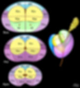
PROSTATE ANATOMY
Four different regions of different composition are recognized in the prostate:
AFS: Anterior fibromuscular stroma
TZ: Transition zone, which surrounds the prostatic urethra. Typically enlarged in men with prostatic hypertrophy, it is what determines the symptoms.
CZ: Central Zone, located at the base of the prostate, posterior to the transition zone, surrounds the right and left ejaculatory ducts.
PZ: Peripheral area, posterior and lateral
75% of tumors originate from the peripheral zone (PZ).
25% of tumors originate from the transition zone (TZ).
A small proportion of tumors originate from the anterior fibromuscular stroma or from the central area.
INTERPRETATION OF IMAGES
The term "multiparametric" derives from the three fundamental parameters of the prostate MRI:
high-resolution axial T2 sequences
the DWI sequences with ADC map
the perfusion study (with the contrast medium)
The comparison of these three parameters allows to categorize the prostatic findings according to the PI-RADS classification, which includes 5 categories, from 1 to 5, with a progressive increase in the probability that the highlighted areas are clinically relevant neoplasms, from very unlikely for the category 1 to very likely for category 5.
The probability of a clinically significant tumor is:
PI-RADS 1 – Very low
PI-RADS 2 – Low
PI-RADS 3 – Intermediate/doubtful
PI-RADS 4 – High
PI-RADS 5 – Very high
The interpretation of the images, and the criteria for assigning the PI-RADS score, are different for the peripheral zone and for the transition zone, and this difference must be duly taken into account during the reporting.


As already mentioned, the definitive diagnosis of prostate cancer is histological, therefore in case of suspicion it is mandatory to perform a prostate biopsy.